
Figure 6.
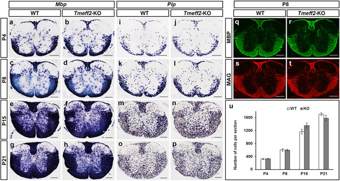
Normal differentiation of OLs in Tmeff2-KO mutant spinal cords. (a–p) Spinal cord sections of wild type and Tmeff2-KO mice were subjected to ISH with Mbp (a–h) and Plp (i–p) riboprobe at different stages. (q–t) Immunofluorescent staining with anti-MBP (q,r) and anti-MAG (s–t) in P8 wild type (q,s) and mutant (r,t) spinal tissues. Bar, 200 μm. (k) Statistical analysis of the number of Plp+ OLs in spinal cords from P4 to P21 stages. n = 3, p > 0.05.