Figure 7.

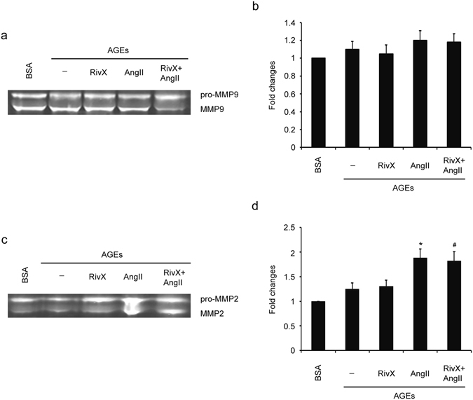
Rivaroxaban did not suppress activities of MMP9 and MMP2 in the presence of AngII in HUVECs. HUVECs were pre-incubated with 100 μg/ml AGE-BSA and non-glycated BSA for 2 h. Cells were then treated with 30 nM rivaroxaban (RivX) or 200 nM AngII for 4 hours. (a) MMP9 activity in the cell culture medium was determined through gelatin zymography analysis. (b) MMP9 activity was expressed as fold changes over that of the BSA control group. The bars show the mean ± SEM (n = 3). (c) MMP2 activity in the cell culture medium was determined through gelatin zymography analysis. (d) MMP2 activity was expressed as fold changes over that of the BSA control group. Cropped images for the gels were shown in (a) and (c). And images for the full-length gels can be found in the supplementary information. The bars show the mean ± SEM (n = 3). * P < 0.05 compared with the parallel blank group, and # P < 0.05 compared with the parallel RivX alone group.
