Figure 3.

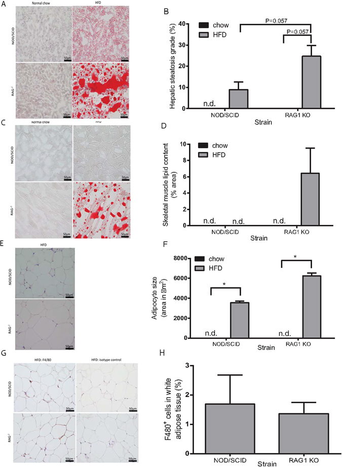
HFD increases lipid storage in Rag1 −/− and NOD/SCID mice with more pronounced effects in Rag1 −/− mice. (A) Oil-red-O stained liver histological sections demonstrate hepatic lipid in NOD/SCID and Rag1 −/− mice fed HFD for 28 weeks. Hepatic lipid is absent in mice fed low-fat chow. (B) Hepatic steatosis (% oil-red-O stained hepatic adipocyte area) in Rag1 −/− and NOD/SCID mice fed HFD. (C) Lipid accumulation in oil-red-O stained skeletal muscle in Rag1 −/− mice fed HFD, but not in Rag1 −/− normal chow-fed mice, or NOD/SCID mice. (D) Intramyocellular lipid content (% oil-red-O stained adipocyte area) in Rag1 −/− mice fed HFD. (E) White adipose tissue deposits (haematoxylin and eosin) in HFD-fed NOD/SCID and Rag1 −/− mice, are absent in mice fed low-fat chow. (F) Mean adipocyte size (expressed as area) is greater in Rag1 −/− mice fed HFD compared to NOD/SCID HFD-fed mice. (G) F4/80 positive (brown) immunostaining of white adipose tissue deposits demonstrates macrophage infiltration in white adipose tissue in NOD/SCID and Rag1 −/− mice fed HFD. (H) Percent of F4/80 positive stained cells in HFD-fed NOD/SCID and Rag1 −/− mice white adipose tissue. Mean + SEM. Kruskal-Wallis and Mann-Whitney test. *P ≤ 0.05. n.d. = not detectable. Scale bar = 50 µm.
