Figure 5.

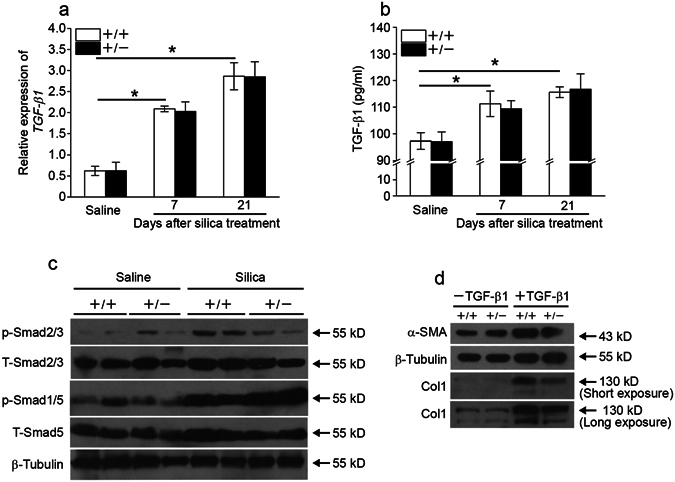
Fstl1 modulates myofibroblast differentiation via facilitating TGF-β1 signaling. (a) qRT-PCR analysis of TGF-β1 mRNA expression in lung tissues of Fstl1 +/− and WT mice at indicated time after saline or silica exposure (n = 4 per group; *P < 0.05 by one-way ANOVA followed by Student’s t test). (b) ELISA analysis of active form of TGF-β1 protein in lung tissues of Fstl1 +/− and WT mice at indicated time after saline or silica exposure (n = 4 per group; *P < 0.05 by one-way ANOVA followed by Student’s t test). (c) The levels of phosphorylation of Smad2/3 (p-Smad2/3), total Smad2/3 (T-Smad2/3), phosphorylation of Smad1/5 (p-Smad1/5) and total Smad1/5 (T-Smad1/5) in lung tissues of of Fstl1 +/− and WT mice 21 days after saline or silica exposure were determined by western blot analysis. β-tubulin was used as a loading control. (d) Primary lung fibroblasts from Fstl1 +/− and WT mice were treated with 5 ng/ml TGF-β1. Protein expressions of α-SMA in cell extracts and type I collagen (Col1) in medium 24 h after TGF-β1 treatment were determined by western blot analysis. β-tubulin was used as a loading control.
