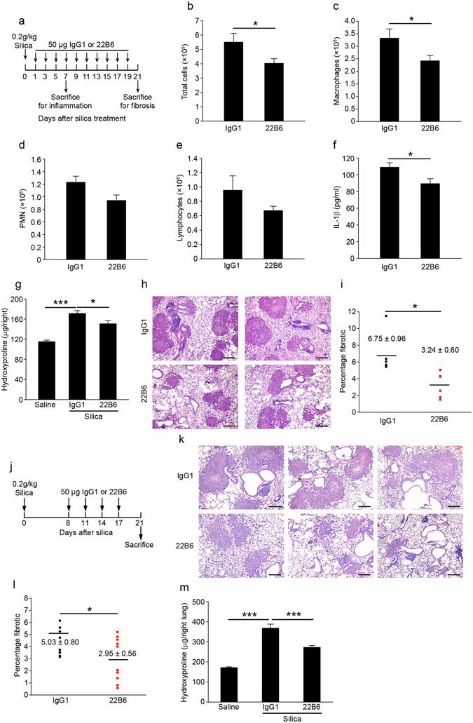
Figure 6.

FSTL1-neutralizing antibody attenuates silica-induced lung inflammation and subsequent pulmonary fibrosis in mice. (a) In a FSTL1 blockage experiment, C57BL/6 mice were intraperitoneally injected with 22B6 mAb or IgG1 (n = 6 per group) every other day from 1 day after silica challenge till the mice were sacrificed on day 7 for inflammation analysis or day 21 for fibrosis analysis. (b–f) For inflammation analysis, (b) the number of total BALF cells was determined by hemocytometer (*P < 0.05 by one-way ANOVA followed by Student’s t test). (c–e) The differential cell counts in BALF were determined according to standard morphologic criteria. (c) Macrophages (*P < 0.05 by one-way ANOVA followed by Student’s t test). (d) PMNs. (e) Lymphocytes. (f) The level of cytokine IL-1β was detected by ELISA assay (*P < 0.05 by one-way ANOVA followed by Student’s t test). (g–i) For fibrosis analysis, (g) hydroxyproline contents in lung tissues were measured (*P < 0.05, ***P < 0.001 by one-way ANOVA followed by Student’s t test). (h) Representative images of the H&E staining of lung sections are shown (Scale bars, 200 μm). (i) Lung fibrotic score analysis of the lung sections. The fibrotic area is presented as a percentage (*P < 0.05 by one-way ANOVA followed by Student’s t test). (j) Interventional dosing regimen of lung fibrosis model. C57BL/6J mice were intraperitoneally injected with 22B6 mAb or IgG1 (n = 10 per group) at indicated time after silica exposure, and lungs were harvested on day 21. (k) Representative images of the H&E staining of lung sections are shown (Scale bars, 200 μm). (l) Lung fibrotic score analysis of the lung sections. The fibrotic area is presented as a percentage (*P < 0.05 by one-way ANOVA followed by Student’s t test). (m) Hydroxyproline contents in lung tissues (***P < 0.001 by one-way ANOVA followed by Student’s t test).
