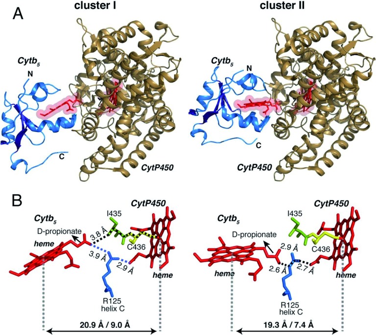
Figure 3. First high-resolution structure of the membrane-bound cytochromes-b5-P450 complex revealing electron transfer pathway.
( A) Three-dimensional structure of the membrane-bound rabbit cytochrome P4502B4–cytochrome b 5 complex reproduced from Ahuja et al. 19. It was obtained by using the high-resolution (solution- and solid-state) nuclear magnetic resonance (NMR) structure of membrane-bound rabbit cytochrome b 5 (PDB code is 2M33), crystal structure of the soluble domain of cytochrome P4502B4 (PDB code is 1SUO), and NMR and mutational constraints to obtain the structure of the membrane-bound complex. ( B) Electron transfer pathway revealed by HARLEM 63 in the complex structure. PDB, Protein Data Bank.

