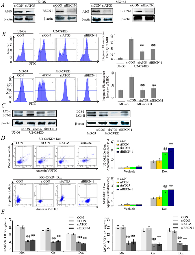
Figure 5.

Inhibition of autophagy restored the chemosensitivity of U2-OS/KD and MG-63/KD cells. (A) Western blot analysis for the expression of BECN-1 and ATG5 proteins. U2-OS/KD cells and MG-63/KD were co-transfected with BECN-1 siRNA (siBECN-1) or ATG5 siRNA (siATG5). After 48 hrs, BECN-1 and ATG5 proteins were detected using Western blot. (B) Flow cytometry assay to detect autophagy level using by MDC staining. Cells were described as (A). (C) Western blot analysis for the expression of LC3-II/I proteins. (D) Flow cytometry for apoptosis analysis using Annexin V-FITC/PI double staining. U2-OS/KD cells and MG-63/KD were co-transfected with siBECN-1 or siATG5. After 48 hrs, the cells were treated with 1 μM Dox for 24 hrs. **P < 0.05 versus U2-OS/KD cells treated with Dox.
