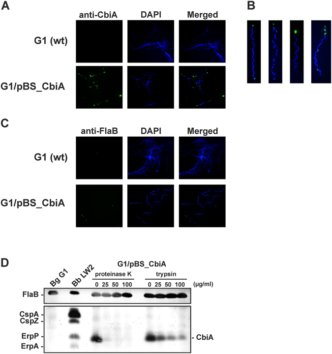
Figure 6.

Surface exposure of CbiA in transformed B. garinii G1. (A) Surface localization of ectopically expressed CbiA was visualized by indirect immunofluorescence microscopy. Spirochetes (6 × 10⁶) were incubated with rabbit anti-CbiA antiserum (1:50) for 1 h at RT with gentle agitation. After fixation, glass slides were incubated with an appropriate AlexaFluor 488-conjugated secondary antibody. For visualization of the spirochetes in a given microscopic field, the DNA-binding dye DAPI was used. The spirochetes were observed at a magnification of 100 × objective. The data were recorded with an Axio Imager M2 fluorescence microscope (Zeiss) equipped with a Spot RT3 camera (Visitron Systems). Each panel shown is representative of at least 20 microscope fields. (B) In situ protease accessibility assay. Native spirochetes were incubated with or without proteinases, then lysed by sonication and total proteins were separated by SDS-PAGE. CbiA was identified by Far Western blot analysis using NHS as source of FH. Flagellin (FlaB) was detected with mAb L41 1C11. FH-binding proteins of B. burgdorferi LW2 (CspA, CspZ, ErpP, ErpA) are indicated on the left and the band corresponding to CbiA on the right. A full-length version is presented in Supplementary Figure S7.
