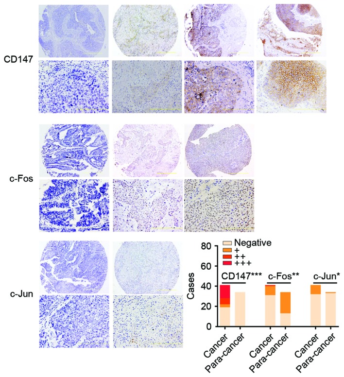
Figure 1.

The immunohistochemical staining of CD147, c-Fos and c-Jun in UCB. The expression of c-Jun, c-Fos and CD147 was scored as negative, absence; +, weak; ++, moderate; and +++, strong. The images were ×100 (upper row) and ×400 magnification (bottom row). The histogram presents the number of cases with CD147, c-Fos and c-Jun expression in UCB tissues and para-cancer normal tissues. The distribution of 41 cases of UCB and 34 cases of para-cancer tissues is presented in the bar graph. CD147 (negative, 19; +, 3; ++, 6; and +++, 13 in UCB. negative, 34 in para-cancer), c-Fos (negative, 31; +, 9; and ++, 1 in UCB. negative, 13; and +, 21 in para-cancer) and c-Jun (negative, 32; +, 9; in UCB. Negative, 33; and +, 1 in para-cancer), respectively. ***P<0.001, **P<0.01 and *P<0.05 vs. para-cancer. UCB, urothelial carcinoma of the bladder.
