Figure 1.

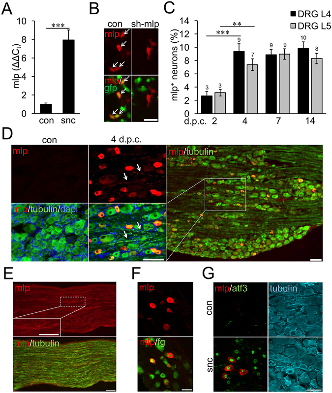
MLP is expressed in a subpopulation of DRG neurons. (A) Quantitative RT-PCR reveals strong induction of Mlp expression in adult male rat L4/L5-DRG 7 days after sciatic nerve crush (snc) in comparison to sham-operated controls (con). Samples of 3 animals per group (snc and con) were used. The Ct values for Mlp were in the range of 24 to 30 cycles and the Ct values for Gapdh of 18–19 cycles. Treatment effect: ***p < 0.001. (B) HEK293 cells were co-transfected with MLP expression plasmid and either scrambled control shRNA (con) or Mlp-shRNA (sh-Mlp). GFP expression indicates shRNA-transfected cells and MLP was detected 24 hours after transfection. Mlp-shRNA knocked down MLP expression. Scale bar: 50 µm. (C) Quantification of MLP-positive (mlp+) L4 and L5 DRG neurons at 2, 4, 7 and 14 days post sciatic nerve crush (d.p.c.). Data represent mean percentages ± SEM of all βIII-tubulin-stained neurons on DRG sections as in (D). Two male rats and 4 sections per DRG were used for each group. Treatment effects: **p < 0.01, ***p < 0.001. (D) Immunohistochemical analysis of MLP expression on DRG sections of sham-operated controls (con) and 4 d.p.c. The sections were stained for MLP (red), βIII-tubulin (green) and DAPI (blue). MLP was detected in a subpopulation of βIII-tubulin-positive neurons and their axons (arrows). Scale bars: 50 µm. (E) MLP expression (red) was also detected in βIII-tubulin-positive axons (green) on proximal sciatic nerve sections at 4 d.p.c. The insert shows a higher magnification of a MLP-positive axon. Scale bars: 100 µm. (F) MLP immunostaining on DRG sections 7 days after sciatic nerve cut and retrograde fluorogold (fg) labeling. MLP expression was only detected in fg-positive injured neurons. Scale bar: 50 µm. (G) DRG sections of sham-operated controls (con) and 4 d.p.c were stained for MLP (red), ATF3 (green) and βIII-tubulin (cyan). ATF3 expression indicated axotomized DRG neurons. MLP was only detected in ATF3-positive neurons. Scale bar: 50 µm.
