
Figure 1.
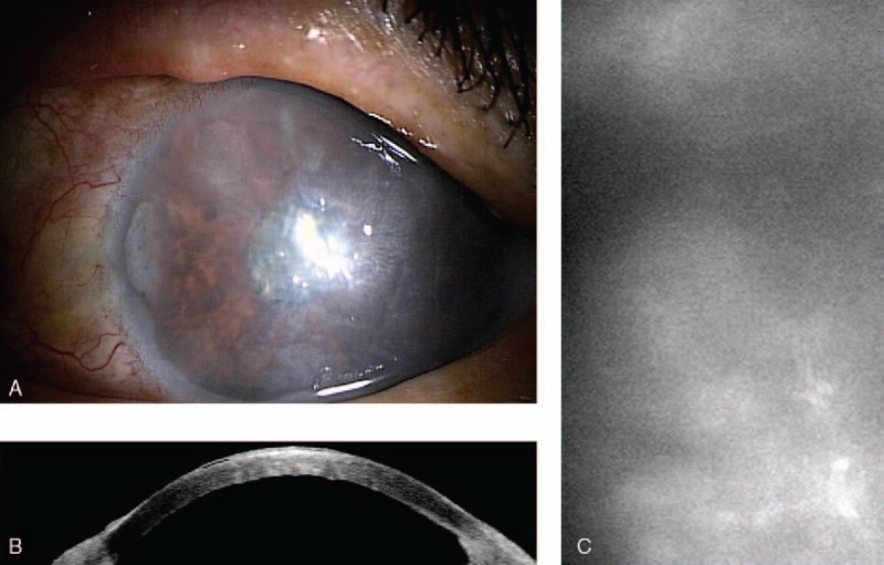
A, Preoperative diffuse bullous keratopathy of left cornea. B, Preoperative anterior ocular coherent tomography of left eye. C, Preoperative left corneal endothelial picture by specular microscopy.
Official websites use .gov
A
.gov website belongs to an official
government organization in the United States.
Secure .gov websites use HTTPS
A lock (
) or https:// means you've safely
connected to the .gov website. Share sensitive
information only on official, secure websites.
A, Preoperative diffuse bullous keratopathy of left cornea. B, Preoperative anterior ocular coherent tomography of left eye. C, Preoperative left corneal endothelial picture by specular microscopy.