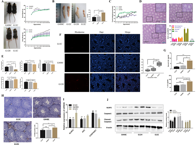
Figure 5.

Influence of a HFD on the reproductive system of mice can be recovered by stopping the HFD. (A) Representative picture of G8C4H, G12H and G12C mice. Comparison of time-dependent increases in bodyweight between G4H8C, G12H and G12C mice (n = 10 in each group). (B) Epididymal adipose tissue and EAT/BW of G4H8C, G12H and G12C mice. (C) Food intake in three groups. (D) Haematoxylin and eosin-stained testicular sections from G4H8C, G12H and G12C mice. Scale bars = 50 μm. (E) Testosterone levels, Spermatic parameters, including sperm concentration, mobility and rate of abnormal sperm, and the number of offspring in each group. (F) Germ cell apoptosis was tested using a TUNEL assay in three groups, and the apoptosis index was determined. (G) Serum and testis homogenates tested for IL1β (ELISA). (H) Immunohistochemical detection of the macrophage-specific antigen F4/80 (black arrows) in testes from G4H8C, G12H and G12C mice and the ratio positive cell between these groups. (I) Inflammasome-related gene expression in these three groups (*P < 0.05 compared with the G12H mice group). (J) Inflammasome protein expression in these three groups. Data are expressed as the mean ± SEM. *P < 0.05, **P < 0.01. ***P < 0.001; n.s. non-significant difference.
