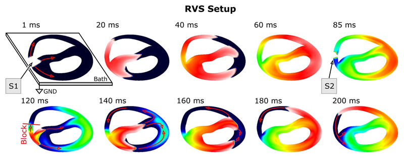
Fig. 1.
Setup used to benchmark sequential simulation runs: a sustained anatomical reentry was induced with an S1–S2 pacing protocol. Shown are polarization patterns of Vm for selected instants. Red arrows indicate the conduction pathways of the activation wavefronts. Due to the presence of a bath, the degrees of freedom associated with the elliptic problem are higher than with the parabolic problem. The left upper panel shows the bath geometry and the location of the reference electrode.

