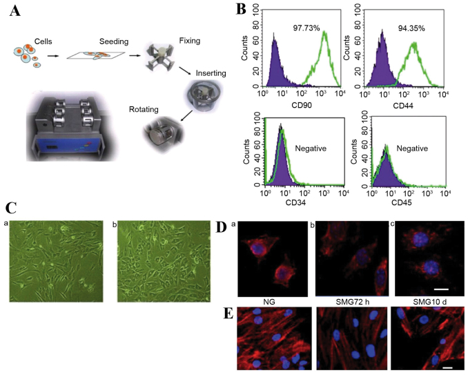
Figure 1.
(A) The SMG clinostat model system. (B) Fluorescence-activated cell sorting analysis of CD44, CD34, CD90 and CD45 in rMSCs. (C) Light microscopy analysis of the shape change of rMSCs under (a) NG and (b) SMG conditions (x50 magnification). (D) Microtubule formation was disrupted following SMG. Scale bar=10 µm. Modified microtubules were observed in rMSCs compared to (a) the NG control following (b) 72 h SMG, but cells appeared to have reestablished microtubules following (c) 10 days SMG. (E) α-actin filaments underwent remodeling following SMG stimulation, appearing diffuse compared to (a) the NG control following (b) 72 h SMG. However, following (c) 10 day SMG, re-concentrated actin was observed. SMG, simulated microgravity; rMSCs, rat mesenchymal stem cells; NG, normal gravity.

