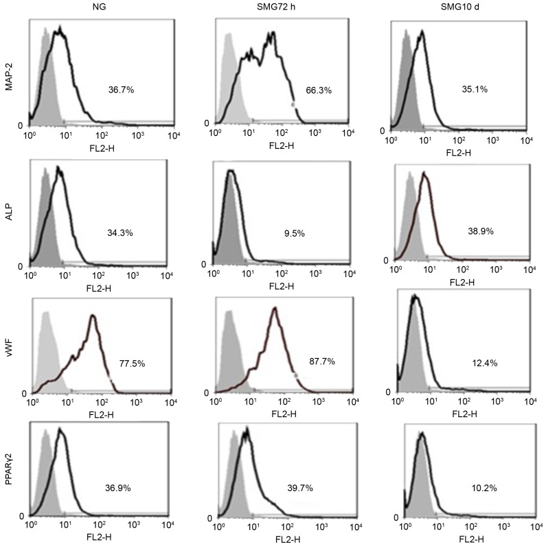
Figure 3.
Fluorescence-activated cell sorting analysis of MAP-2, vWF, ALP and PPARγ-2 following culture of rat mesenchymal stem cells for 10 days in differentiation medium subsequent to exposure to NG and 72 h and 10 days SMG. The gray area is the isotype control. The results represent three independent experiments. MAP-2, microtubule-associated protein 2; vWF, von Willebrand factor; ALP, alkaline phosphatase; PPARγ2, peroxisome proliferator-activated receptor γ2; NG, normal gravity; SMG, simulated microgravity.

