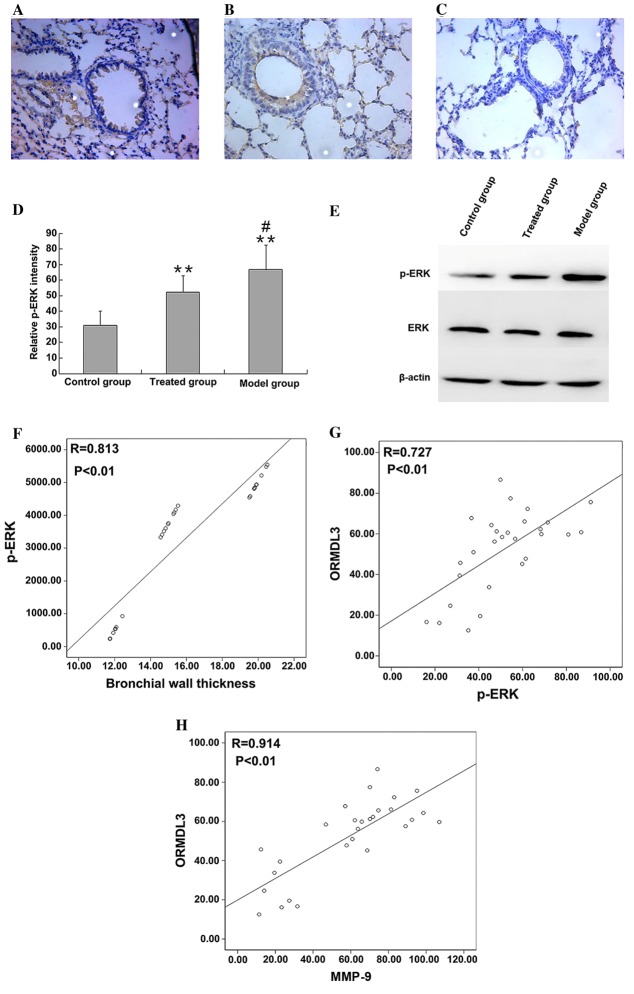
Figure 5.
Expression levels of p-ERK protein in lung tissues of the (A) asthmatic model (B) budesonide-treated and (C) healthy control groups were investigated using immunohistochemistry. Magnification, ×100. (D) p-ERK protein expression was quantified using ImageJ analysis of stained sections and (E) western blotting. RNA expression level of MMP-9 in lung tissues was determined using reverse transcription-quantitative polymerase chain reaction. **P<0.01 vs. control; #P<0.05 vs. treated. (F) p-ERK protein expression was positively associated with bronchial wall thickness (r=0.813, P<0.01) and (G) ORMDL3 protein expression was positively associated with p-ERK level (r=0.727, P<0.01). (H) MMP-9 protein expression was positively associated with ORMDL3 expression level (r=0.914, P<0.01). p-ERK, phosphorylated-extracellular-signal regulated kinase; ORMDL3, orosomucoid-like 3; MMP-9, matrix metalloproteinase 9.

