
Figure 3.
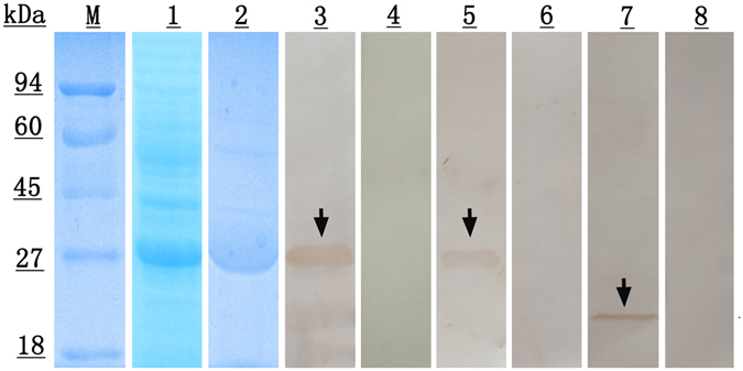
SDS-PAGE and western blotting analysis of Eg-DHFR. M, protein molecular weight markers; lane 1, E. coli BL21 (DE3) lysate from IPTG-induced cells expressing rEg-DHFR; lane 2, purified rEg-DHFR; lane 3, purified rEg-DHFR probed with anti-rEg-DHFR rabbit serum; lane 4, purified rEg-DHFR probed with native (preimmune) rabbit serum; lane 5, purified rEg-DHFR probed with the serum of E. granulosus infected sheep; lane 6, purified rEg-DHFR probed with native (healthy) sheep serum; lane 7, the total protein from protoscoleces probed with anti-Eg-DHFR rabbit serum; lane 8, the total protein from protoscoleces probed with native (preimmune) rabbit serum.