
Figure 4.
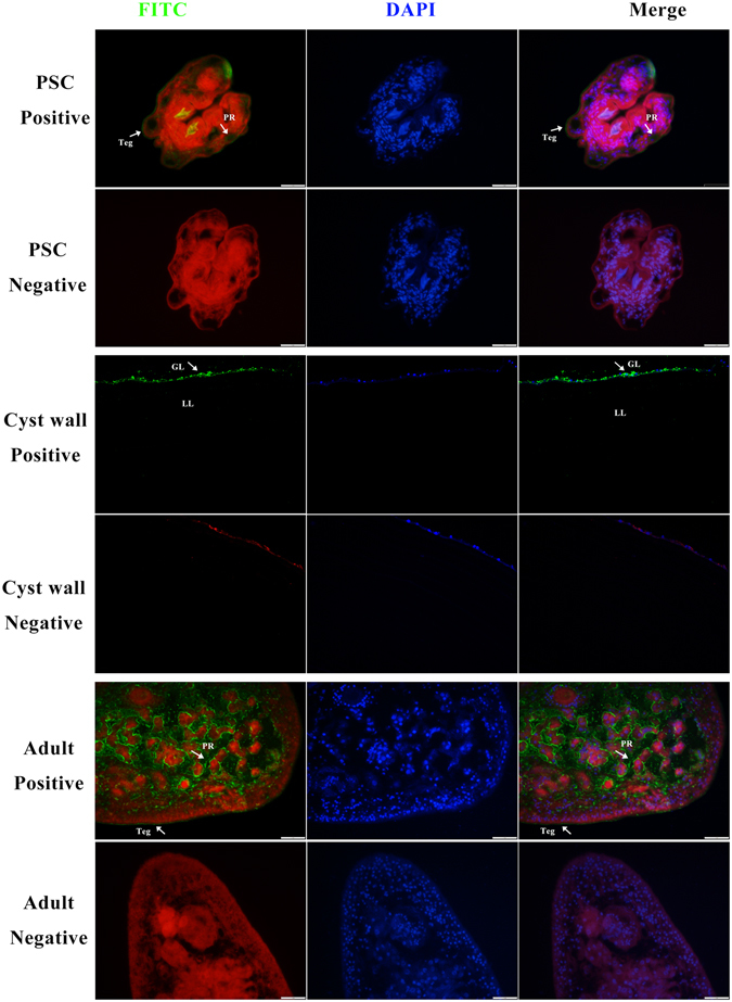
Immunofluorescent localisation of Eg-DHFR in different stages of E. granulosus. Eg-DHFR was localised in the protoscolex, germinal layer and adult worm using specific anti-rEg-DHFR IgG (positive), or preimmune serum (negative). The nucleus DNA was stained with DAPI (blue). Abbreviations: Teg, tegument; PR, parenchymal region; GL, germinal layer; LL, laminated layer. Scale bars: 1 mm.