Figure 5.

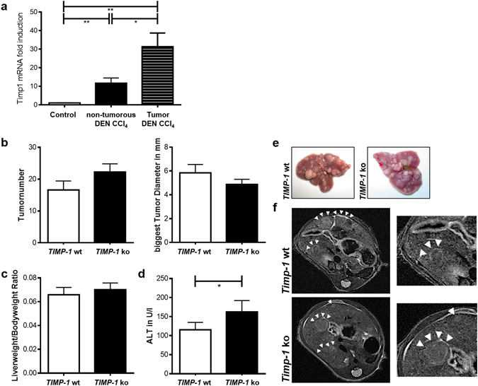
TIMP-1 deficient mice are not protected from fibrosis-driven hepatic carcinogenesis. (a) TIMP-1 expression increased in HCC tissue compared to adjacent paired normal tissue and control liver tissue following juvenile DEN administration and chronic CCl4 injection. (b) Tumorload does not differ between TIMP-1 wt and ko as assessed by tumor number and size and (e) visualized by representative pictures of excised livers and (f) in vivo MRI images. (c) TIMP-1-deficiency does not alter the liverweight/bodyweight ratio. (d) A combination of juvenile DEN administration and chronic CCl4 treatment led to overall increased ALT level, whereas elevated level could be measured in serum from TIMP-1 ko mice as compared with TIMP-1 wt. Bar columns represent mean ± standard error of the mean. **p < 0.01; *p < 0.05. (n = 13–19).
