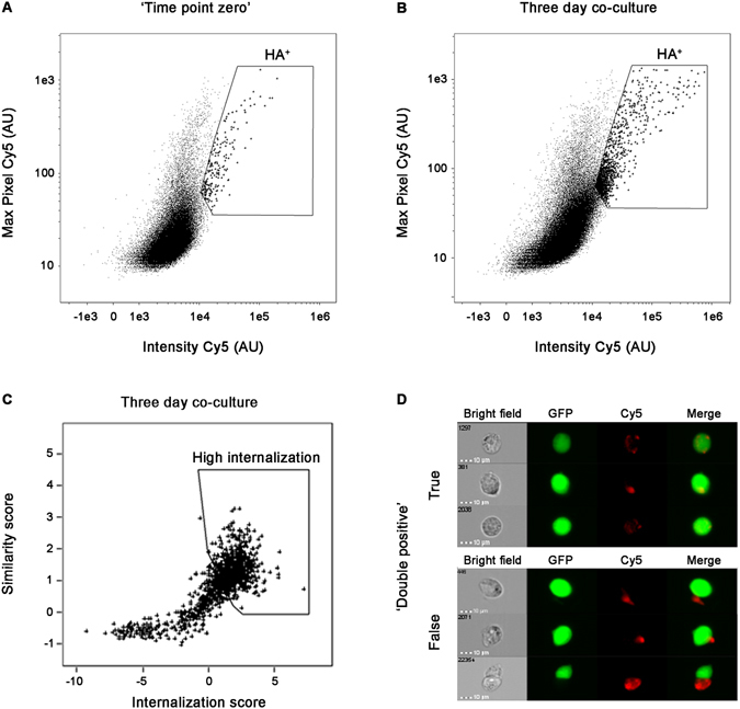
Figure 3.

IFC analysis of GFP-expressing ‘recipient’ cells co-cultured with ‘donor’ cells expressing HA-tagged α-synuclein. (A,B) Bivariate analysis of GFP-expressing ‘recipient’ cells collected from (A) ‘time point zero’ and (B) three day co-culture. HA+ gated cells are demarcated. (C) Double positive (HA+) cells collected after three days co-culture were further gated to exclude Cy5 staining outside cell boundaries using the Internalization score and the Similarity score between the GFP and Cy5 channels (see Materials and Methods for details). The gated cells represent a high Internalization score within the GFP-expressing cells. (D) Images representing true and false ‘double positive’ cells.
