
Figure 5.
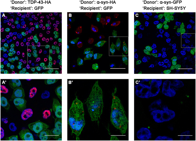
Transmission of tagged TDP-43 and α-synuclein into ‘recipient’ cells. The boxed area in (A,B,C) is enlarged in (A’,B’,C’), respectively. GFP-expressing ‘recipient’ cells were co-cultured with either HA-tagged TDP-43 (A,A’) or α-synuclein (B,B’) ‘donor’ cells for three days and viewed using confocal microscopy. Alternatively, ‘donor’ cells expressing GFP-tagged α-synuclein were co-cultured with naïve SH-SY5Y ‘recipient’ cells (C,C’). DAPI (blue), anti-HA-antibody (red). Scale bars: (A) (50 µm); (A’) (20 µm); (B,C) (40 µm); (B’) (12 µm); (C’) (8 µm).