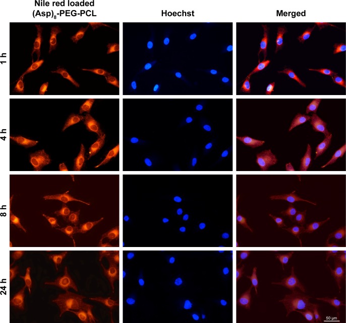
Figure 6.
Fluorescence microscopic images of MG63 cells incubated with Nile red-loaded (Asp)8-PEG-PCL nanoparticles at a concentration of 100 μg/mL at 37°C. Cell nuclei were counterstained using Hoechst blue, and the blue and red fields of fluorescence images were merged at each time point.
Abbreviation: (Asp)8-PEG-PCL, polyaspartic acid peptides-poly (ethylene glycol)-poly (ε-caprolactone) polymer.

