Fig. 3.
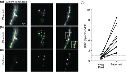
Contrast improvement with intensity gradient holographic illumination. (a) Fluorescence images of small sections of basal dendrites from three L5 pyramidal neurons under wide-field illumination obtained by a large homogeneous CGH pattern covering the whole field of view. Scale bar: 1 μm. (b) Scheme of the multispot CGH illumination patterns superimposed on the wide-field fluorescence image. (c) VSD fluorescence under CGH-patterned illumination. (d) Increase in contrast expressed as the fluorescence intensity ratio between spines and parent dendrites as determined in nine different preparations. The dramatic increase in the intensity ratio by a factor of 2 to 14 corresponds to an equivalent decrease in the contamination of the spine signal by scattered light from the parent dendrite.

