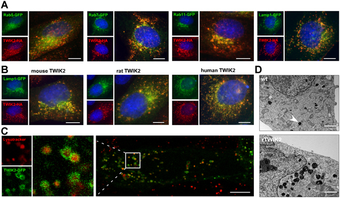
Figure 2.

Localization of TWIK2 in lysosomal membranes. Representative images of MDCK cells expressing (A) GFP-coupled compartment markers and rat TWIK2HA, and (B) Lamp1-GFP together with mouse, rat or human TWIK2HA. TWIK2 was stained with HA antibody (red) and nuclei were visualized with Hoechst33342 (blue). Scale bar: 10 µm. (C) Image of a living MDCK cell expressing rat TWIK2-GFP and incubated with lysotracker (red), staining the lysosomal lumen. Scale bar: 10 µm. (D) Electronic microscopy images of a cell expressing rat TWIK2 (lower panel) or of a control cell (upper panel). Scale bar: 2 µm. Lysosomes are dense bodies (plain arrow).
