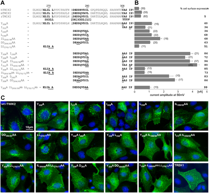
Figure 4.

Identification of trafficking motifs in the C-ter of TWIK2. (A) Sequence of human, rat and mouse TWIK2 C-ter. The trafficking motifs are shown in bold. Overview of the set of generated mutants, in which underlined amino acids were replaced by an alanine residue. (B) Current amplitudes at +50 mV of the corresponding rat TWIK2 mutants recorded in Xenopus oocytes (number of recorded oocytes) expressed as mean current amplitude ± sem. Mutant rat TWIK2HA channels were transfected into MDCK cells and stained with HA antibody. % cell surface expression corresponds to the percentage of transfected cells showing a clear expression of TWIK2 at the plasma membrane. (C) Representative images of MDCK cells expressing the different TWIK2 mutations shown in (B) and TREK1. Scale bar: 10 µm.
