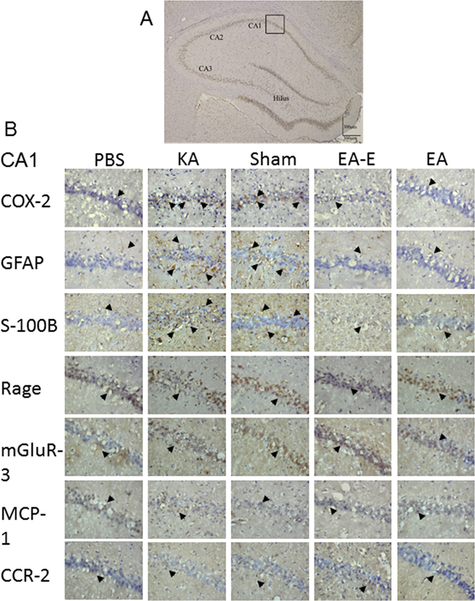
Figure 3.

Effects of electric stimulation at the ear and electro-acupuncture at ST36–ST37 on the counts of COX-2, GFAP, S100-B, RAGE, mGluR3, MCP-1, and CCR2 immunoreactive cells in kainic acid (KA)-induced epileptic seizure rats. The CA1, CA2, CA3, and hilus regions of the hippocampus (A). The counts of COX-2, GFAP, and S100-B immunoreactive cells were higher in the KA group (KA) and sham group (Sham) than in the normal group (Normal), whereas these counts were decreased in the auricular group (Ear) and Zusanli group (Zusanli). The counts of RAGE, mGluR3, MCP-1, and CCR2 immunoreactive cells were similar among the normal, KA, sham, and auricular groups (B). Immunoreactive cell (arrowhead); B image is 400× in the CA1 region; n = 6.
