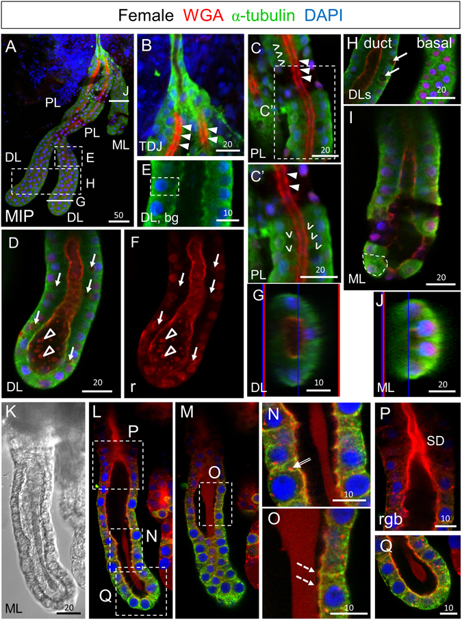
Figure 1.

SGs from newly eclosed adult females comprise cuboidal secretory cells and a central WGA-positive maturing apical duct. (A) Low magnification maximum intensity “Z” projection (MIP) confocal image of a SG about 30 minutes post eclosion stained with antibodies to newly synthesized alpha-tubulin (green; tubulin antibody) and the dyes WGA (red; O-GlcNAcylated proteins and chitin) and DAPI (blue; nuclei). (B) Confocal image of the proximal triductal junction (TDJ) region showing high levels of alpha-tubulin in the cell bodies of duct cells and high levels of periodic WGA staining in the apical duct region of the most proximal secretory cells. Arrowheads indicate lower intensity staining near cell junctions. (C) Confocal image of the proximal lobe (PL) with variable levels of alpha-tubulin staining of cuboidal-shaped cells and high-level periodic apical WGA staining in the duct region. Arrowheads indicate lower intensity staining near cell junctions. Carets mark sites of secretory cavity formation. (C’) Zoom image of (C). (D–F) Single confocal images of the distal lobe (DL) showing cuboidal shaped cells (dashed outline, E) with high levels of WGA staining at the apical surface as well as lower and particulate WGA staining in the lumen (open arrowheads). Arrows (D,F) indicate pre-apical compartments (PACs). (G) Cross section of the DL reveals cuboidal alpha-tubulin positive secretory cells with apical WGA staining surrounding the central lumen. The thin blue and red lines (G,J) are positional markers added by the “Ortho” function in the Zen software. (H) Confocal images of DLs showing a central (duct) section (left) or a basal section (right). Arrows indicate PACs. (I,J) Sagittal and cross sections of the medial lobe with variable staining of alpha-tubulin and low level apical WGA staining. The dashed line (I) highlights the cuboidal shape of the cells. (K–Q) Early ML showing faint WGA staining of the apical matrix. (K) DIC image of ML. (L,M) Different focal planes of same gland showing the apical matrix either pulled away from or contacting the cuboidal secretory cells. (N) WGA-positive vesicles are observed within secretory cells of the ML (double-lined arrow). (O) The apical matrix directly contacts the apical domains of the secretory cells in some areas (dashed arrows). (P,Q) Zoomed images of (L). Individual channels are denoted “b”–blue, DAPI, “g”–green, tubulin, or “r”–red, WGA. Scale bar lengths are in microns. No primary antibody control imaging is shown in Supplementary Figure S1.
