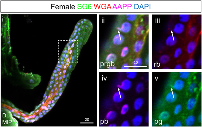
Figure 6.

Small pre-apical compartments are located near the nucleus and presumptive Golgi prior to expansion. Shown are confocal images from a female SG DL lobe around 12 hours post eclosion. Apical WGA staining is weak through most of the DL lobe (i). Some nuclei show small accumulations of secretory proteins and weak WGA in the absence of DNA, near the nucleus and presumptive Golgi (ii–v, arrow). These may be sites of pre-apical compartment formation. Individual channels are denoted “p”, “b”, “g”, or “r”. “MIP” images are maximum intensity Z-projections. Scale bar lengths are given in microns.
