Figure 6.

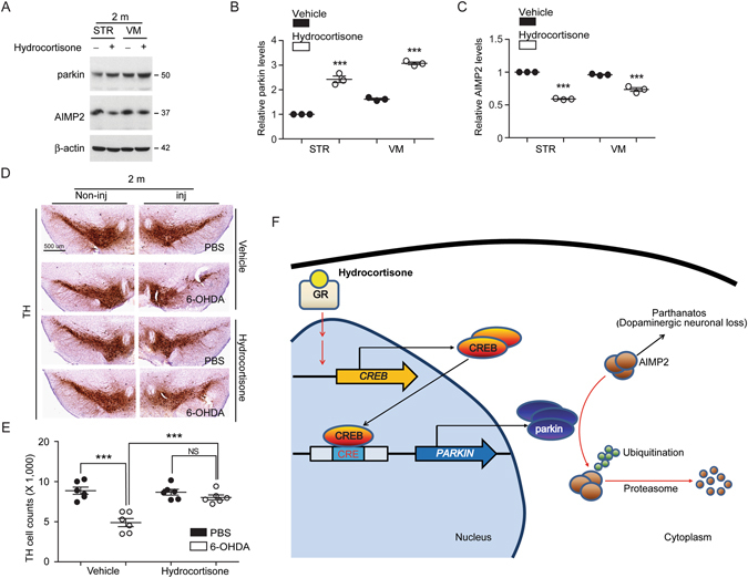
Hydrocortisone prevents dopamine cell loss in 6-OHDA mouse model. (A,B,C) Western blot analysis of parkin and its substrate AIMP2 in the striatum and ventral midbrain regions of mice (2 months old) treated with hydrocortisone or DMSO for 7 days. Relative parkin or AIMP2 protein levels were normalized to that of β-actin and shown as plot graphs (n = 3 mice per group). (D) Representative tyrosine hydroxylase (TH) immunohistochemistry (scale bar = 500 um) of the substantia nigra of 6-OHDA PD mouse models treated with hydrocortisone or DMSO. 6-OHDA (8 µg) was stereotaxically injected into the striatum (coordinate from bregma, L: −2.0, AP: 0.5, DV: −3.0 mm) to model dopaminergic neurodegeneration in mice. (E) Stereological assessment of tyrosine hydroxylase (TH)-positive dopaminergic neurons in the substantia nigra pars compacta of injection side from the indicated mouse groups (n = 6 mice per group). (F) Schematic summary of proposed molecular pathways illustrating hydrocortisone-mediated induction of parkin expression. Hydrocortisone binds to glucocorticoid receptor which in turn leads to expression of CREB. CREB increases parkin expression via binding to CREB binding motifs of parkin promoter region. Hydrocortisone-stimulated parkin expression results in the downregulation of the toxic parkin substrate AIMP2, which is beneficial for dopaminergic neuronal survival. Quantified data are expressed as mean ± s.e.m. ***P < 0.001, unpaired two-tailed Student’s t test (B,C) and ANOVA test followed by Tukey post hoc analysis (E). Full blots (for cropped images in A) are presented in Figure S6.
