Figure 1.

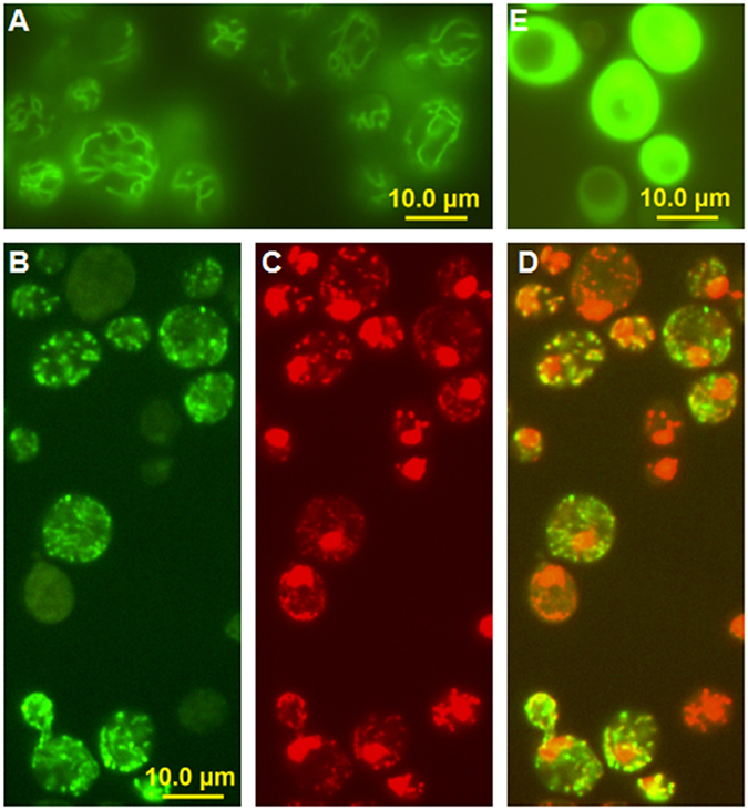
Fluorescence microscopy showing the mitochondrial localization of yeast Lon protease. (A) The mitochondrial network in Saccharomyces cerevisiae cells after DiOC6 staining. (B) The localization of Lon-GFP expressed from pUG35 in S. cerevisiae. (C) Visualization of S. cerevisiae nuclear (large red dots) and mitochondrial DNA (smaller red spots) after DAPI staining. The blue colour of DAPI was changed to red in the imaging software. (D) The merged signal confirming the co-localization of Lon-GFP and mtDNA. (E) A negative control, showing the non-specific GFP signal from an empty pUG35 plasmid which localizes to the cytosol of S. cerevisiae cells.
