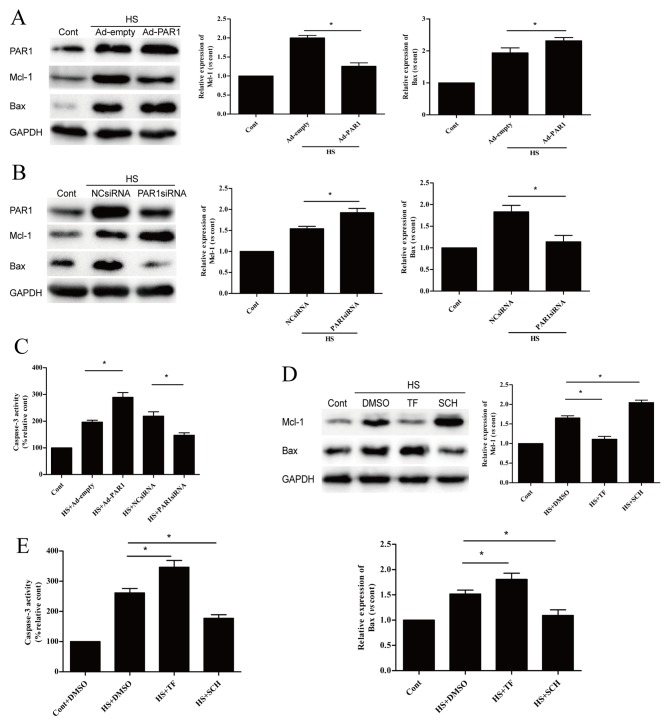
Figure 3.
Effects of PAR1 on apoptosis-associated proteins following heat stress in HUVECs. Protein expression levels of Mcl-1 and Bax were detected by western blot analysis following transfection with (A) Ad-empty or Ad-PAR1, or (B) NC siRNA or PAR1 siRNA for >48 h. HUVECs were incubated at 37°C (control) or 43°C (heat stress) for 90 min, followed by a 6-h recovery period at 37°C. (C) Caspase-3 enzymatic activity was measured in the cell lysates. Untransfected cells were pretreated with DMSO or 40 µM TF for 10 min or 150 nM SCH for 1 h prior to incubation at 37°C (control) or 43°C (heat stress) for 90 min, followed by a 6-h recovery period at 37°C. (D) Mcl-1 and Bax proteins were identified by western blotting analysis. (E) Enzymatic activity of caspase-3 was measured in the cell lysates. Data are presented as the mean ± standard deviation of three independent experiments. *P<0.05; **P<0.01; ***P<0.001. PAR1, protease-activated receptor 1; HUVECs, human umbilical vein endothelial cells; Ad, adenovirus; NC, negative control; siRNA, small interfering RNA; Mcl-1, myeloid cell leukemia 1; Bax, B-cell lymphoma 2 associated X; TF, TFLLR-NH2; SCH, SCH79797; DMSO, dimethyl sulfoxide; HS, heat stress; Cont, control.

