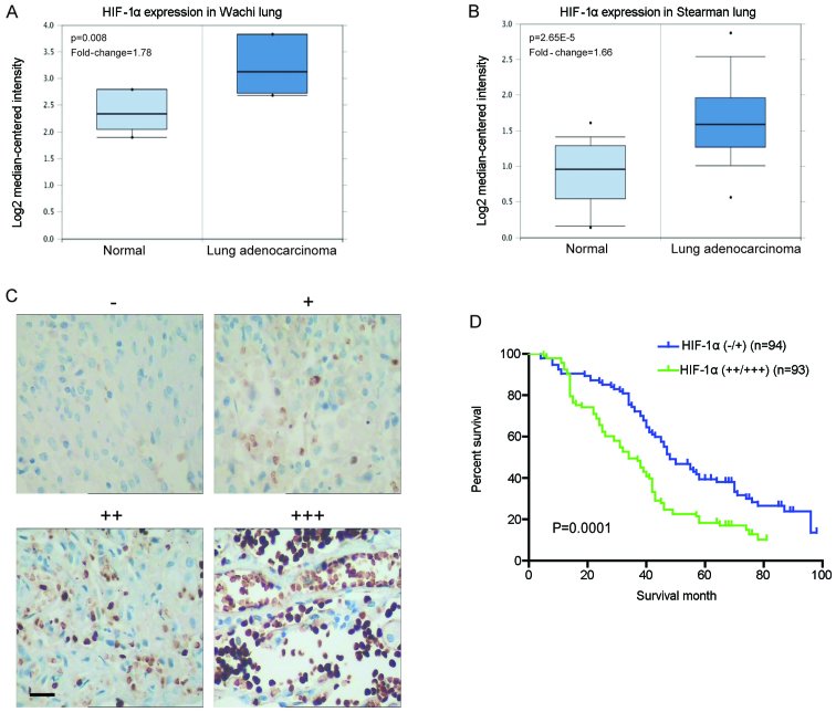
Figure 1.
Aberrant high expression of hypoxia-inducible factor 1α (HIF-1α) in clinical non-small cell lung cancer tissues indicates poor patient prognosis. (A and B) Investigation of lung cancer microarray data sets using Oncomine (www.oncomine.org). (A) Higher HIF-1α mRNA levels were found in squamous cell lung carcinoma (n=5) when compared with normal lung tissues (n=5) in the Wachi lung data set. (B) Higher HIF-1α mRNA levels were found in lung adenocarcinoma (n=20) compared with normal lung tissues (n=19) in the Stearman lung data set. (C) The expression level of HIF-1α protein in clinical specimens of lung cancer was detected using immunohistochemistry with respect to the HIF-1α expression intensity: negative (−), positive (+), strongly positive (++) and very strongly positive (+++) immunoreactivity (scale bar, 50 µm). (D) The association of HIF-1α expression with the prognosis of lung cancer patients. Kaplan-Meier overall survival curves indicated that low expression of HIF-1α in lung cancer tissues was significantly associated with a better overall survival rate. ‘Low’ or ‘high’ HIF-1α level was defined according to its cut-off value, which was defined as the median value of the cohort of patients tested; P<0.05 by log-rank test.

