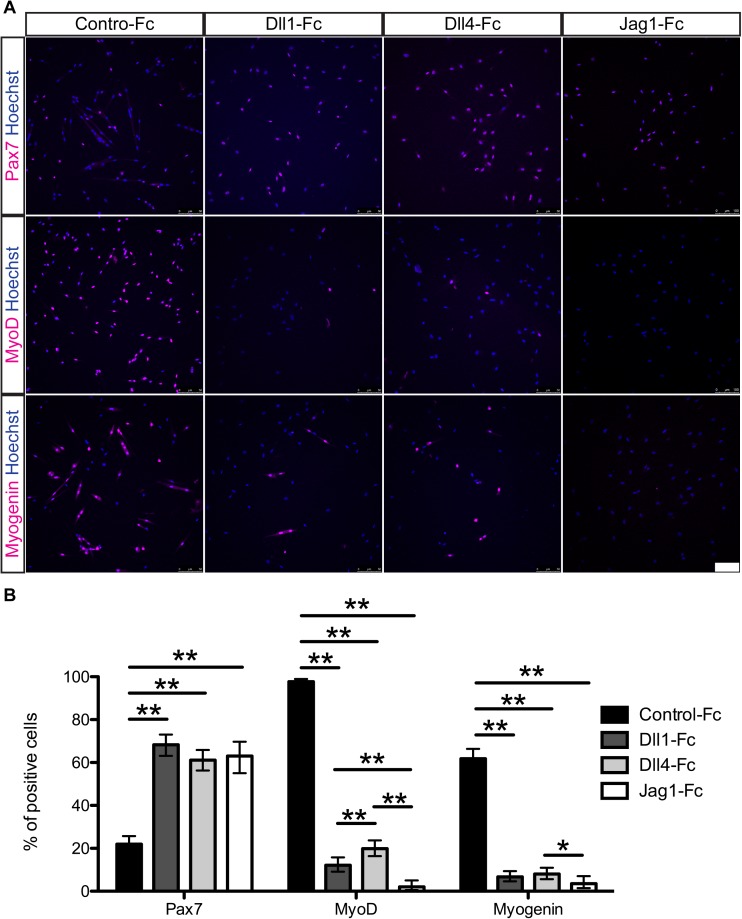
Fig 2. Dll1-Fc, Dll4-Fc, and Jag1-Fc retain Pax7 expression but repress activation markers in mouse myogenic cells ex vivo.
(A) Immunocytochemistry for Pax7, MyoD, and Myogenin of mouse myogenic cells cultured with or without Notch ligands. Hoechst staining reveals nuclei. Scale bar = 100 μm. (B) Quantitative analysis of Pax7, MyoD, and Myogenin in the cultured cells. Data are reported as mean and 95% confidence interval of 200–450 cells per staining condition. Statistical significance was assessed by 2-sample test for equality of proportions with Holm method. P-value are <0.05 (*) or <0.01 (**).

