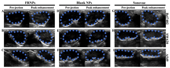
Figure 5.
Imaging results of folate-receptor targeted nanoparticles (FRNPs), blank NPs and Sonovue in the 3 types of xenografts before injection and at the peak enhancement. (A, D and G) Changes in AOI after injection of FRNPs to the 3 types of xenografts under B-mode ultrasonography. (B, E and H) Corresponding changes with the injection of blank NPs. (C, F and I) Corresponding changes with injection of Sonovue. In all images, the areas circled by blue dotted lines represent the areas of interest (AOI) of tumor xenografts.

