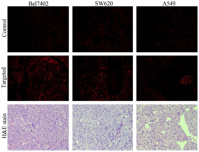
Figure 9.
Fluorescent microscopy images of tumor slices from the xenografts of target and control groups after intravenous injection of folate-receptor targeted nanoparticles (FRNPs) dyed with Cy7 and physiological saline, which observed with 40X objective and ×10 ocular lens. In the targeted group brighter red fluorescent spots, which corresponded to FRNPs dyed with Cy7 in the tumor tissue, were observed with a long-pass 515–560 nm emission filter cube under lamp illumination. The images of H&E-stained tumor slices were observed under an optical microscope.

