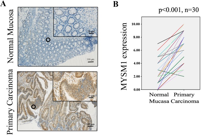
Fig 1. Immunohistochemical staining for MYSM1 in human colorectal tissues.
(A) Expression levels of MYSM1 in normal mucosa (n = 30) and primary carcinoma tissues (n = 30)from patients with CRC were detected by immunohistochemical staining (×10, 100 μm; ×40, 20 μm).(B)MYSM1 protein levels in normal mucosa and primary carcinoma tissues were quantified as shown as line chart.

