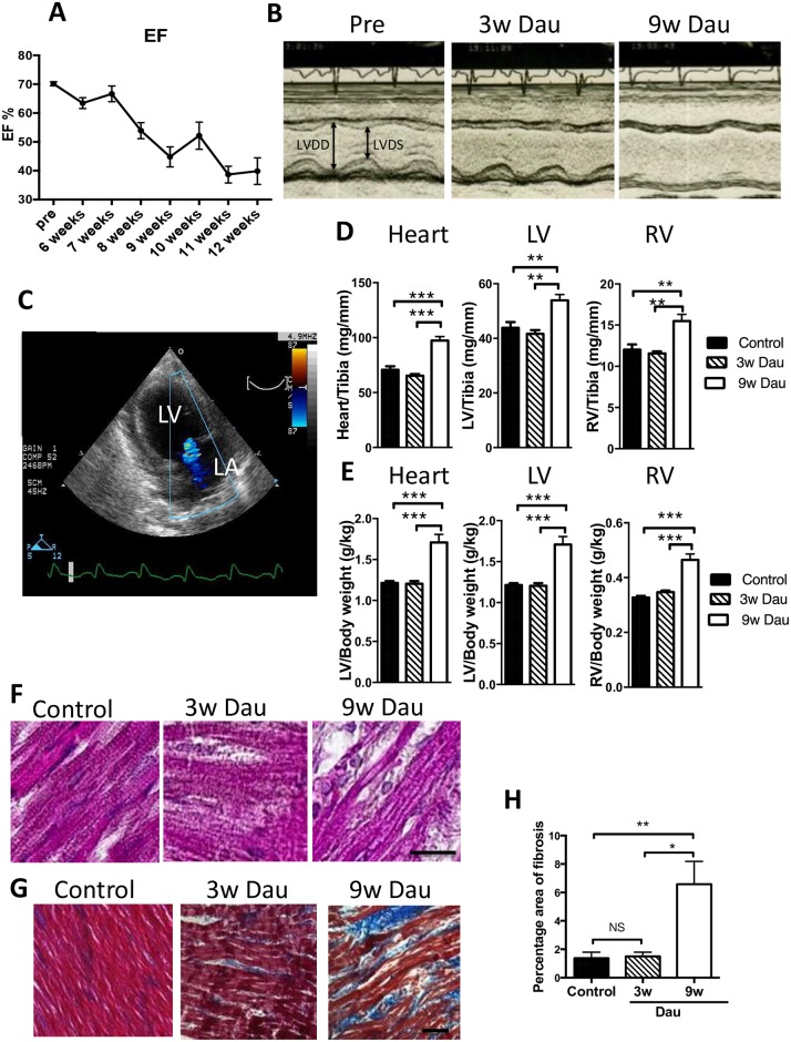
Fig 1. Dau-induced cardiotoxicity in vivo.
(A) Ejection fraction (EF) was decreased with 9w Dau treatment. (n = 8) (B) Representative M-mode image of echocardiography before Dau treatment (Pre) and after 3w and 9w of Dau treatment. (C) Mitral regurgitation was found in the 9w Dau-treated rabbits. LA indicates left atrium. (D) Heart to tibia length ratios for Dau-treated and control rabbits (n = 6–8) (E) Heart to body weight (Bw) ratios for Dau-treated and control rabbits (n = 6–8) (F) Hematoxylin and eosin stain of LV from Dau-treated and control hearts (scale bar = 20μm) (G) Masson trichome stain of LV from Dau-treated and control hearts. Blue indicates fibrosis. (scale bar = 50μm) (H) Quantification of (G) (n = 6–8, respectively), *p<0.05, **p<0.01, ***p<0.001, LV, Left Ventricle; RV, Right Ventricle; LVDS and LVDD, LV dimensions in systole and diastole.

