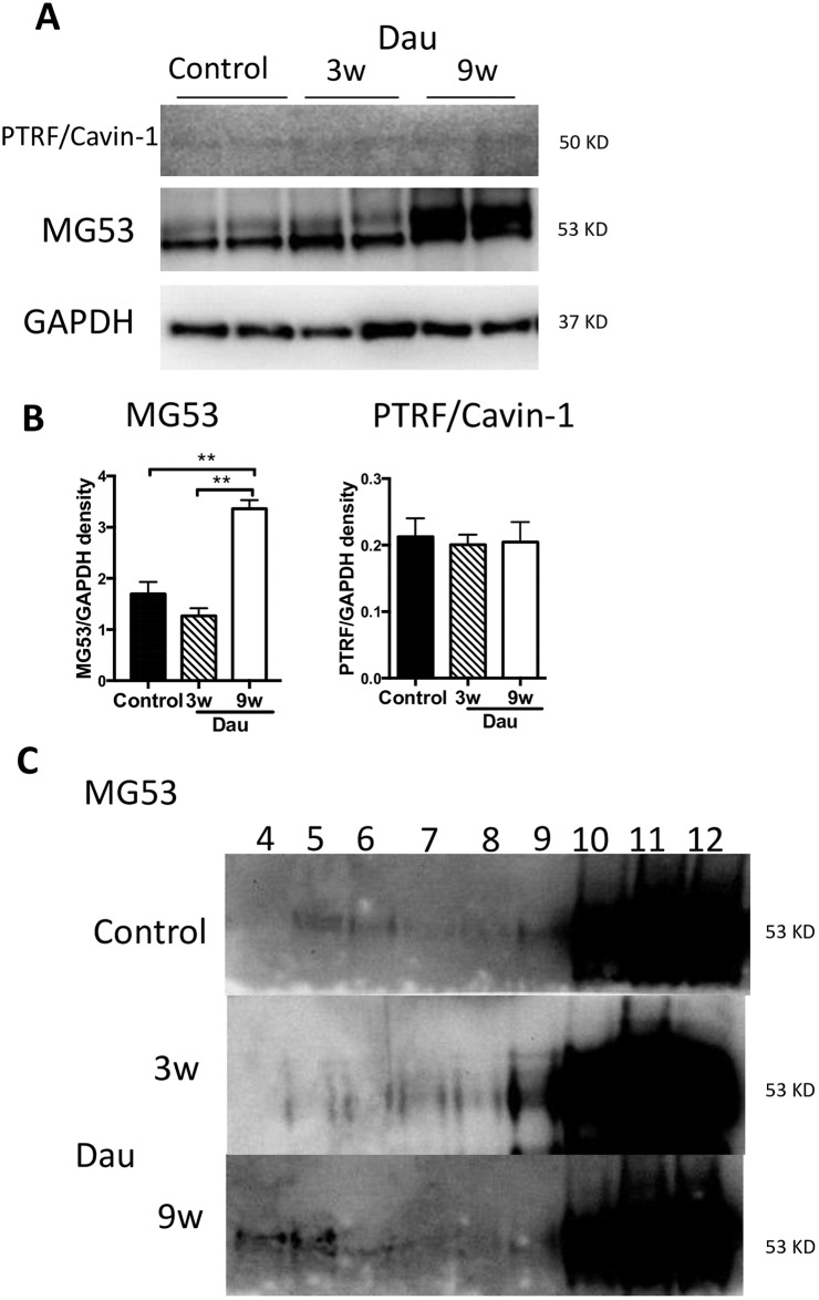
Fig 7. Membrane repair proteins in the Dau-treated heart.
(A) Western blot of MG53, PTRF/cavin-1 in rabbit left ventricle (LV) from Dau-treated and control heart. (B) Quantification of (A), n = 4–6, respectively. Expression of MG53 in the 9w Dau-treated LV was higher than that of control LV. n = 6, respectively (C) Whole heart protein homogenates were biochemically fractionated by sucrose gradient fractionation. MG53 was found in buoyant as well as non-buoyant fractions in LV from Dau-treated and control rabbits. *p<0.01, NS, not significant; MG53, Mitsugumin-53; GAPDH, Glyceraldehyde 3-phosphate dehydrogenase.

