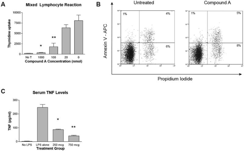
Figure 1. Compound A impairs Tcon proliferation in a mixed lymphocyte reaction and inhibits TNF production in vivo in response to LPS challenge.

(A) 5×10⁴ CD4+CD25− B6 responder cells were incubated with 5×10⁴ irradiated TCD B6D2 stimulator splenocytes. During the last 16–20 hours of incubation 0.037 MBq (1 μCi) of [3H]thymidine was added to each well, and [3H]thymidine incorporation measured by scintillation counting. *P=0.0044 for comparison between 1000nM and 0 Compound A groups by Student’s t test. **P=0.0162 for comparison between 100nM and 0 Compound A groups. (B) 1×10⁶ purified B6 CD4+CD25− Tcons were cultured for 48 hours in T cell media supplemented with murine IL-2 at 100 IUs/ml in the presence or absence of Compound A at 100nM. The cells were then collected and stained for flow cytometry with anti-Annexin V antibody and Propidium Iodide. Cell cultures were done in triplicate and representative flow cytometry plots for each culture condition are depicted. (C) Mice were administered saline or varying amounts of Compound A by IP injection. One hour later mice were given saline (No LPS group) or 200mcg of LPS by IP injection. Two hours later, the mice were killed and blood was collected by cardiac puncture. Serum TNF levels were then measured by ELISA. *P=0.0017 for comparison between 250mcg Compound A and LPS alone groups by Student’s t test. **P=0.0007 for comparison between 750mcg Compound A and LPS alone groups.
