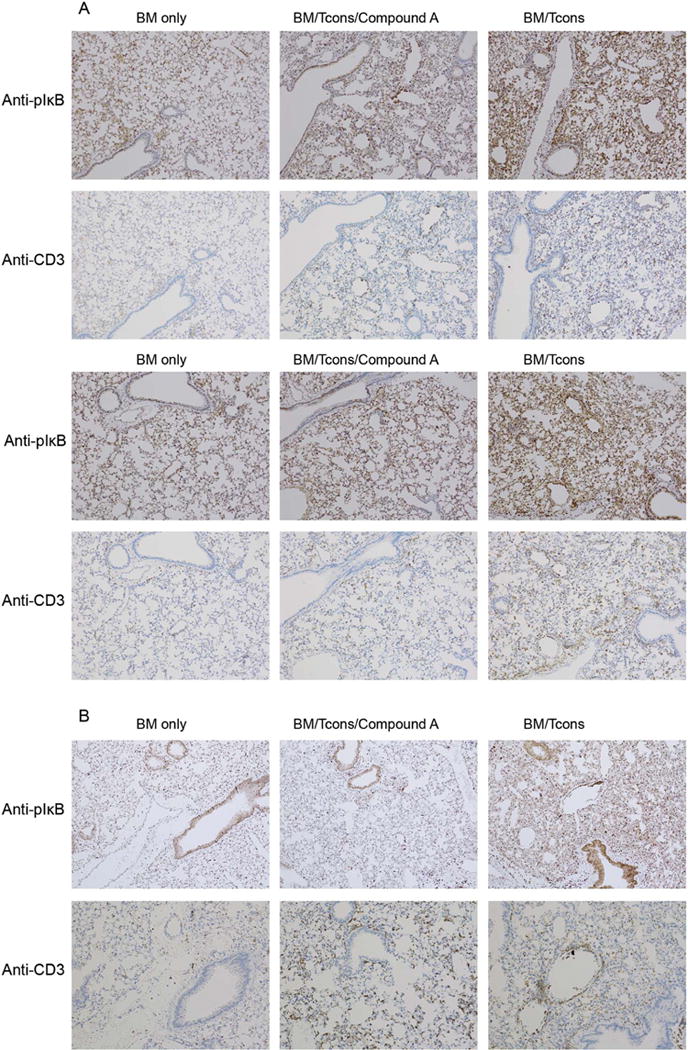
Figure 4. In a murine model of IPS recipient lungs demonstrate diffuse NF-κB signaling for at least three weeks post-transplant and this activity is attenuated by Compound A administration.

(A–B) B10.BR mice were administered cyclophosphamide at 120mg/kg by IP injection on transplant days −3 and −2. They were then irradiated to 700 rads on day −1 on a cesium irradiator. On day 0, recipient mice were administered 1×10⁷ TCD BM cells from B6 mice +/− 7×10⁶ NK1.1-depleted B6 splenocytes +/− Compound A dosed at 175mcg once daily by IP injection on days 0–6. (A) On day +7 the mice were killed and their hearts perfused through with saline. Their lungs were then removed and placed in formalin. The lungs were subsequently sectioned and stained for IHC using an anti-phospho-IκBα antibody. Consecutive slides were also stained with an anti-CD3 antibody. Two mice from each treatment group are depicted. (B) A third set of mice was transplanted similarly but administered daily Compound A on days 0–20. They were then killed on day +21 for IHC (B).
