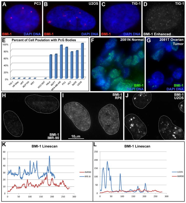
Fig 2. BMI-1 aggregates in large aberrant foci, forming bodies.
A–B) BMI-1 protein accumulates into bodies in cancer cells. C–D) Normal fibroblasts only have faint punctate nucleoplasmic signal. Red channel enhanced at right. Exposures the same for A–C. E) Cancer cell lines contain BMI-1 bodies while non-cancer lines do not. F) Normal human tissue with uniform punctate distribution of BMI-1. G) The matched tumor sample contains BMI-1bodies. H–J) Low nucleoplasmic BMI-1 in fibroblasts (H), slightly higher levels in telomerase immortalized RPE cells (I), and large accumulations in cancer cells (J), with concomitant reduction in nucleoplasmic signal. (H–J same scale). K–L) BMI-1 linescans of the same cells in (H–J).

