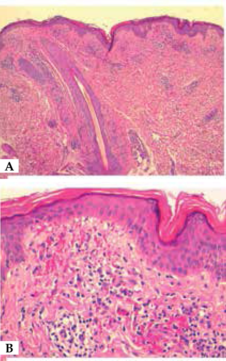
Figure 2.
A. HE 40x: Skin with mild epithelial hyperplasia, occasional foci of spongiosis, and predominantly perivascular superficial lymphohistiocytic infiltrate with extravasation of red blood cells; B. HE 200x: Detail of superficial lymphohistiocytic infiltrate with extravasation of red blood cells

