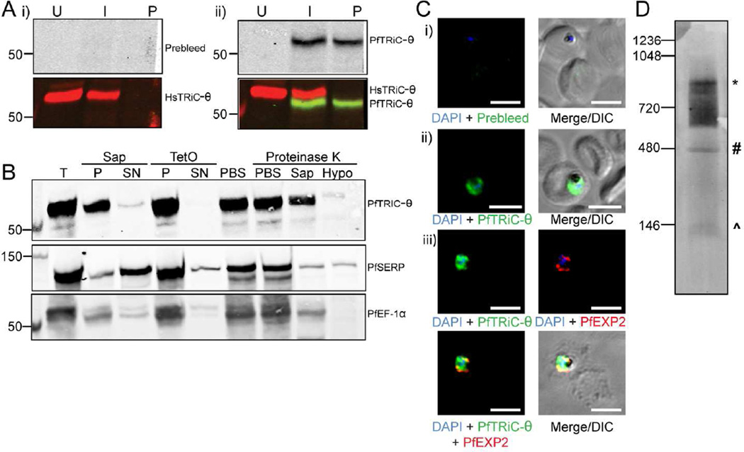
Figure 2. PfTRiC-θ forms a high molecular weight complex in the parasite cytosolic fraction.
A, Lysates from uninfected RBC (U), trophozoite-stage infected RBC (I) or saponin-isolated parasites (P) were extracted with RIPA buffer and probed with antibodies against human TRiC-θ and i) prebleed and ii) immune sera generated against PfTRiC-θ. B, Infected RBC samples (T = total sample) were fractionated with 0.05 % saponin (Sap; 20 s, room temperature) or 1 HU of TetO (20 min, 37 °C). PfSERP is a soluble parasitophorous vacuole protein, released into the saponin-soluble fraction (SN= supernatant), but remaining in the TetO-insoluble fraction (P = pellet). PfEF-1α is a soluble parasite protein, remaining in the saponin- and TetO-insoluble fractions. A proteinase K accessibility assay (last four lanes of blot) was performed with TetO-insoluble fractions from trophozoite-stage infected RBC. The sample was divided into four and incubated in PBS (two samples), 0.05 % saponin, or a hypotonic lysis solution (Hypo). Samples were treated with 1 mg/mL Proteinase K (30 min, on ice) as indicated. Immunoblots, indicating that the protease gains access to PfTRiC-θ after hypotonic lysis, are representative of two independent experiments. C, Immunofluorescence assay (IFA) of paraformaldehyde-fixed (i and ii) or acetone-fixed (iii) trophozoite-infected RBC. DAPI stains the parasite nuclei and PfEXP2 delineates the parasite PV compartment. DIC = differential interference contrast. Scale bars = 5 µm. Data are representative of three independent assays. D, Saponin-isolated parasites were permeabilized with digitonin, the soluble fraction was separated by blue-native polyacrylamide gel electrophoresis and the blot was probed with anti-PfTRiC-θ. * = expected size of the heterohexadecamer. # = expected size of heterooctamer. ^ = expected size of monomers. Immunoblots are representative of three independent assays. Equivalent fractions were loaded in all blots. Marker bands are in kDa.

