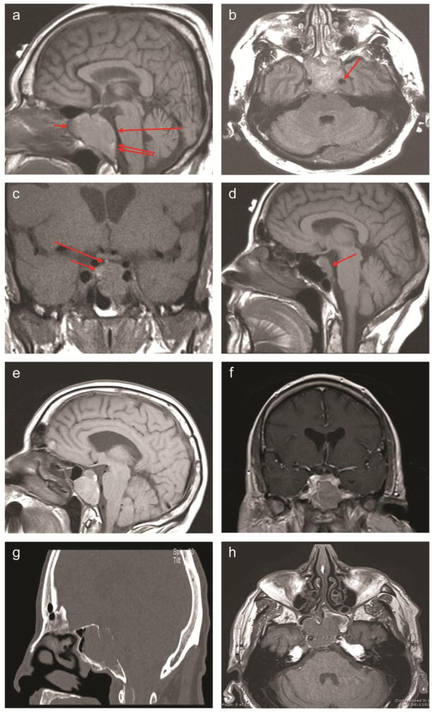
Figure 1. Imaging of parasellar plasmacytomas.
A. Case 1: T1-weighted sagittal image, without contrast. Large mass is seen in the sella and sphenoid sinus (short arrow), expanding the clivus (double arrow) and flattening the ventral belly of the pons (long arrow).
B. Case 1: Flair axial image, without contrast. Large sellar and sphenoid mass is extending into the left cavernous sinus and encasing the internal carotid artery (arrow).
C. Case 2: T1-weighted coronal image, without contrast. Large sellar mass with inferior extension (long arrow) is noted. The infundibulum angles toward presumed residual normal pituitary tissue (short arrow).
D. Case 3: T1-weighted sagittal image, without contrast. Diffusely enlarged pituitary is seen with downward extension into the clivus (arrow).
E, F. Case 4: Sagittal pre-gadolinium (E) and coronal post-gadolinium (F) T1 images. A heterogeneously poorly enhancing T1 hyperintense mass lesion centered within the clivus is noted, with expansion of the clivus and extension into the sphenoid sinuses. A portion of the floor of the sella is absent and there is an intrasellar component of the enhancing mass. The mass invades the bilateral cavernous sinuses.
G, H. Case 5: Sagittal CT (G) and noncontrast T1 axial MRI (H) images. CT shows a large lytic lesion within the clivus with extension into the sphenoid sinus through the posterior wall the sphenoid sinus. There is thinning of the posterior cortex of the clivus with bowing of the mass posteriorly. MRI shows a 33 × 34 × 29 mm hypointense T1 and T2 lesion centered in the clivus, with extension into the sphenoid sinus and likely right cavernous sinus extension.

