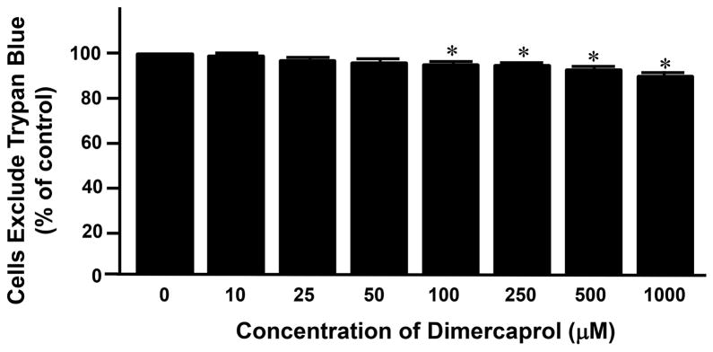
Figure 1.
Cytotoxicity of dimercaprol was tested by Trypan Blue assay. PC-12 cells were exposed to different concentrations of dimercaprol for 4 hrs. Cell viability was determined by trypan blue dye exclusion assay. Viable cells were the cells that excluded the dye. The cell viability of control (dimercaprol at 0 μM) was considered 100%. The viability of cell of other groups was expressed as the percentages of the control. It appears that dimercaprol began to induce significant cell death when its concentration was at and higher than 100 μM (* p<0.05, ANOVA). All data were expressed as mean ± SEM, n=5.

