
Fig. 1.
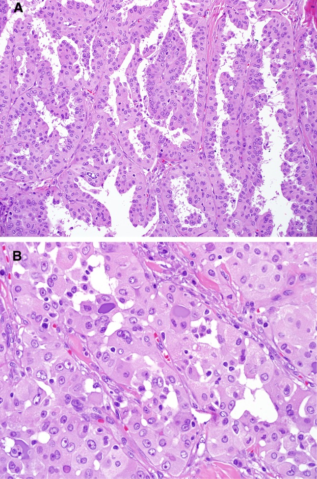
Case 1. The tumor showed focally papillary features with well-defined fibrovascular cores (a). Areas with glandular pattern were admixed with the papillary patterned areas. Areas with the “classic” pattern of MASC were also seen; these areas show dense eosinophilic to purple mucin. The nuclei showed prominent red nucleoli and open clear chromatin (b)