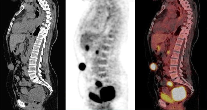
Fig. 1.
Sagittal CT, FDG-PET and fused images of a 37-year-old woman with HIV seropositive serum. She was referred for restaging of cervical adenocarcinoma. CT was non-conclusive about recurrence or residual disease. PET demonstrated metabolically active disease in the pelvis and the presence of a pelvic lymph node. In addition, metabolically active disease noted in the umbilical region due to uptake in the umbilicus associated to peritoneal carcinomatois (atypical finding). This finding was confirmed by biopsy. A follow-up scan 6 months later (not shown) revealed a rapidly progressive disease

