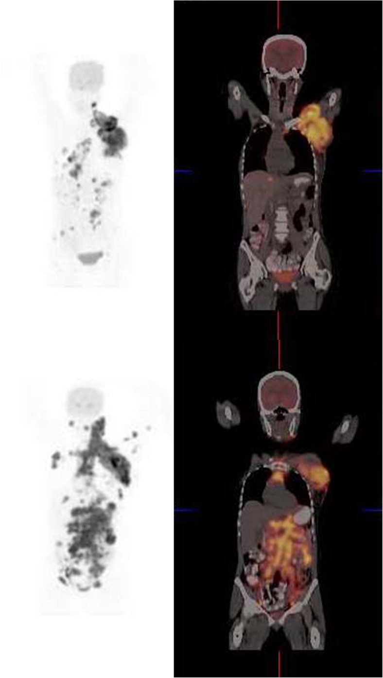
Fig. 3.
Coronal FDG-PET and fusion images of a 41-year-old woman with HIV infection and diffuse large B-cell lymphoma diagnosed by biopsy of a large mass in the left axilla. She has been on HAART for 17 months. Her CD4 count was 354 and her viral load less than 50 copies per ml. The scan shows FDG-avid lymph nodes in the left axilla, hilar, para-aortic, abdominal and pelvic nodes. There is also pelvic bone involvement. Lymphadenopathy is due to DLBL and not reactive lymph nodes of HIV when a scan is interpreted along with immunological parameters. On follow-up there was rapid progression of disease

