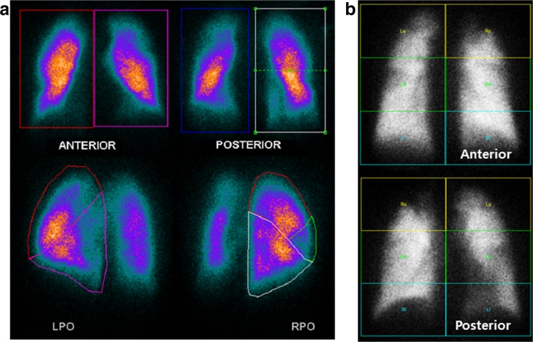
Fig. 1.
Planar lung scan based segmentation methods. (a) PO method: each lung was divided into lobar regions from the posterior oblique view image. The counts of each lobe were divided by the total counts over the ipsilateral lung measured from the same oblique projection. This fraction was then multiplied by the fractional contribution of the ipsilateral lung, obtained from the anterior and posterior projections, (b) AP method: three equal rectangular regions of interest (ROI) on anterior and posterior views were drawn: top, middle, and bottom. The counts in each ROI were divided by the total counts over the ipsilateral lung measured from the anterior and posterior projections

