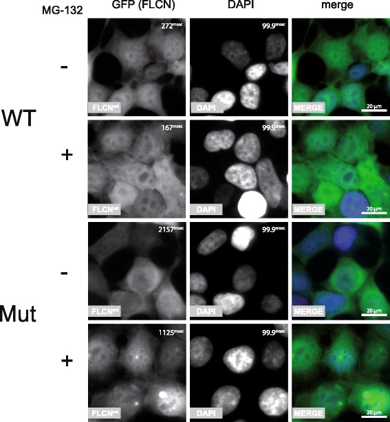
Fig. 6.

HEK293T cells expressing FLCN WT or Mut fused to GFP using the TALEN technology were subjected to immunofluorescent imaging. WT FLCN localizes to both cytoplasm and nucleus, the mutant protein is primarily found in the cytoplasm. Interestingly treatment with MG-132 did not only stabilize the mutant protein which is reflected by a shorter exposure time during image acquisition but also led to a nuclear shift making its localization more similar to the WT protein (bar = 20 μm, exposure time of the GFP and DAPI channel are depicted in millisecond in the upper right corner of each panel)
